Kręgi lędźwiowe
Kręgi lędźwiowe (łac. vertebrae lumbales, skrót: L) – kręgi w lędźwiowym odcinku kręgosłupa. U człowieka jest ich 5 (L1 - L5), u koni, bydła, jelenia szlachetnego (Cervus elaphus) i kozy[1] 6, u owcy i świni 6–7, natomiast u psa i kota 7[2]. Trzony kręgów lędźwiowych są stosunkowo długie, o spłaszczonych głowach i dołach. Wyrostki poprzeczne przypominają tu cienkie blaszki określane jako wyrostki żebrowe. Na wyrostkach stawowych doczaszkowych obecne są wyrostki suteczkowate[1]. U ptaków kręgi lędźwiowe są zrośnięte i wchodzą w część synsakrum[3].
Przypisy
- ↑ a b Helena Przespolewska, Henryk Kobryń, Tomasz Szara & Bartłomiej J. Bartyzel: Podstawy anatomii zwierząt domowych. Warszawa: PWN, 2014, s. 12–14. ISBN 978-83-62815-22-7.
- ↑ Simon Hillson: Mammal Bones and Teeth: An Introductory Guide to Methods of Identification. Routledge, 2016, s. 25. ISBN 978-1-315-42500-9.
- ↑ III Szkielet. W: Bronisław Ferens i Roman J. Wojtusiak: Ornitologia ogólna. Ptak, jego budowa i życie. Warszawa: Państwowe Wydawnictwo Naukowe, 1960, s. 73–74.
Galeria

Kręg lędźwiowy człowieka
Kręgi lędźwiowe człowieka

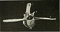
Kręg lędźwiowy kota
Kręg lędźwiowy konia

Synsakrum strusia (5), którego częścią są kręgi lędźwiowe
![]() Przeczytaj ostrzeżenie dotyczące informacji medycznych i pokrewnych zamieszczonych w Wikipedii.
Przeczytaj ostrzeżenie dotyczące informacji medycznych i pokrewnych zamieszczonych w Wikipedii.
Media użyte na tej stronie

The Star of Life, medical symbol used on some ambulances.
Star of Life was designed/created by a National Highway Traffic Safety Administration (US Gov) employee and is thus in the public domain._(20398618288).jpg)
Autor: Internet Archive Book Images, Licencja: No restrictions
Title: The cat : an introduction to the study of backboned animals, especially mammals
Identifier: catintroduction00miva (find matches)
Year: 1881 (1880s)
Authors: Mivart, St. George Jackson, 1827-1900
Subjects: Cats; Anatomy, Comparative
Publisher: New York : Scribner's
Contributing Library: The Library of Congress
Digitizing Sponsor: The Library of Congress
View Book Page: Book Viewer
About This Book: Catalog Entry
View All Images: All Images From Book
Click here to view book online to see this illustration in context in a browseable online version of this book.
Text Appearing Before Image:
40 THE CAT. chap. m. posterior dorsal vertebrae, the first lumbar being quite like the ast dorsal, except that it has no capitular surface, but, in its place, a short forwardly extending transverse process, and that the niet- apophyses are somewhat larger. As we proceed backwards through the series of lumbar vertebrae,
Text Appearing After Image:
Fig. 18.—Fifth Lumbar Vertebra. a. Anapophysis. c. Centrum. to. Metapophysis. n. Neural lamina. s. Neural spine. t. Transverse process. z. Prezygapophysis. 3. Postzygapophysis. the anapophysis decreases, so that in the sixth lumbar there is but a minute rudiment of such a process. The metapophysis is at its maximum in the fourth lumbar vertebra, but is large even in the last. The neural spine is longest at the fourth. The transverse process increases rapidly from the first lumbar vertebra to the fourth, and is slightly longer in the fifth and sixth lumbar vertebrae. The zygapophyses continue to be directed as in the fifth lumbar vertebra, except that the postzygapophyses of the seventh look once again more downwards. The centrum of the seventh lumbar vertebra is not longer than is that of the first, and the same is the case with the neural arch. § 8. Having noted the characters of the vertebrae next behind the dorsal ones, we may advance to those in front of them. Of the seven cervical vertebrjb the first two are sufficiently exceptional to demand separate notice. The other cervicals are very much alike, but the fifth may be selected for comparison with the fifth dorsal vertebra. Its centrum is relatively wider from side to side and narrower
Note About Images

Autor: Anatomography, Licencja: CC BY-SA 2.1 jp
Lumbar vertebrae (shown in red).
_(14585562440).jpg)
Autor: Internet Archive Book Images, Licencja: No restrictions
Lumbar vertebra of horse
Identifier: horseitstreatm05axej (find matches)
Title: The horse, its treatment in health and disease with a complete guide to breeding, training and management
Year: 1906 (1900s)
Authors: Axe, J. Wortley
Subjects: Horses
Publisher: London, Gresham Pub. Co.
Contributing Library: NCSU Libraries
Digitizing Sponsor: NCSU Libraries
View Book Page: Book Viewer
About This Book: Catalog Entry
View All Images: All Images From Book
Click here to view book online to see this illustration in context in a browseable online version of this book.
Text Appearing Before Image:
Fig. 284.- -Dorsal Vertebra (Front\iew) Superior Spinous Process.- Transverse Process. Articula-tion for Tubercle of Rib. •■ Articu-lation for Head of Rib. 5 ..\ntoriorArticular Face of Body. •* SpinalCanal. 181 HKALTH AM) DISEASE cesses, which are directed horizontally outwards. The last two are muchthicker and somewhat shorter than the rest, and are united to each other
Text Appearing After Image:
Fig. 285.—Lumbar Vertcl.r.. 1 Superior Spinous Proce.ss. - Anterior Oblique Process. Transverse Process. •* AnteriorArticular Face of Body. ° Spinal Canal. l)v the borders of their transverse processes, and to the transverse processof the sacrum by synovial articulations. The Sacrum (fig. 28G) or rump bone, as we have already pointed
Note About Images

Star of life, blue version. Represents the Rod of Asclepius, with a snake around it, on a 6-branch star shaped as the cross of 3 thick 3:1 rectangles.
Design:
The logo is basically unicolor, most often a slate or medium blue, but this design uses a slightly lighter shade of blue for the outer outline of the cross, and the outlines of the rod and of the snake. The background is transparent (but the star includes a small inner plain white outline). This makes this image usable and visible on any background, including blue. The light shade of color for the outlines makes the form more visible at smaller resolutions, so that the image can easily be used as an icon.
This SVG file was manually created to specify alignments, to use only integers at the core 192x192 size, to get smooth curves on connection points (without any angle), to make a perfect logo centered in a exact square, to use a more precise geometry for the star and to use slate blue color with slightly lighter outlines on the cross, the rod and snake.
Finally, the SVG file is clean and contains no unnecessary XML elements or attributes, CSS styles or transforms that are usually added silently by common SVG editors (like Sodipodi or Inkscape) and that just pollute the final document, so it just needs the core SVG elements for the rendering. This is why its file size is so small.
A lumbar vertebra from above and behind.

Autor: Autor nie został podany w rozpoznawalny automatycznie sposób. Założono, że to Uwe Gille (w oparciu o szablon praw autorskich)., Licencja: CC-BY-SA-3.0
pelvis and synsacrum of an ostrich, ventral view
1 Os pubis, 2 Os ischii, 3 Os ilium, 4 symphysis pubica, 5 Synsacrum, 6 Fossae renales, 7 Os femoris, 8 Pygostyl