Pleśniak biały
| ||
 Plecha pleśniaka białego | ||
| Systematyka | ||
| Domena | eukarionty | |
| Królestwo | grzyby | |
| Gromada | Mucoromycota | |
| Klasa | Mucoromycetes | |
| Rząd | pleśniakowce | |
| Rodzina | pleśniakowate | |
| Rodzaj | pleśniak | |
| Gatunek | pleśniak biały | |
| Nazwa systematyczna | ||
| Mucor mucedo Fresen. Beitr. Mykol. 1: 7 (1850) | ||
Pleśniak biały (Mucor mucedo Fresen.) – gatunek grzybów należący do rodziny pleśniakowatych (Mucoraceae)[1].
Systematyka i nazewnictwo
Pozycja w klasyfikacji według Index Fungorum: Mucor, Mucoraceae, Mucorales, Incertae sedis, Mucoromycetes, Mucoromycotina, Mucoromycota, Fungi[1].
Niektóre synonimy nazwy naukowej[2]:
- Mucor brevipes Riess 1853
- Mucor coprophilus Povah 1917
- Mucor dependens (Burgeff) Burgeff 1924
- Mucor griseo-ochraceus Naumov 1915
- Mucor murorum Naumov 1915
- Mucor proliferus Schostak. 1896
- Mucor rigidus L. Léger 1895
- Mucor sphaerocephalus Bull. 1791
- Mucor vulgaris P. Micheli 1729
Rozmnażanie
Jest komórczakiem, to znaczy, że zbudowany jest z jednej, ale wielojądrowej komórki tworzącej strzępkę przerastającą podłoże. Ze strzępki tej wyrastają wysokie trzonki zarodnionośne zwane sporangioforami. Na ich szczycie powstają kuliste, czarne zarodnie (sporangia), w których wytwarzane są bezpłciowo zarodniki (sporangiospory). Są one haploidalne i zróżnicowane płciowo na dwa typy, oznaczane zazwyczaj jako (+) i (-). Po pęknięciu zarodni zarodniki wysypują się i mogą być roznoszone przez wiatr, wodę lub zwierzęta. W sprzyjających warunkach kiełkują wytwarzając rozgałęzione, wielojądrowe strzępki o haploidalnej liczbie chromosomów (cenocyty). Strzępki te morfologicznie nie różnią się one od siebie, są jednak zróżnicowane płciowo. Zarówno strzępki (+), jak i strzępki i (-) mogą wytwarzać zarodnie[3].
Gdy zetkną się dwie odmienne płciowo strzępki (+) i (-), zachodzi między nimi rozmnażanie płciowe. Najpierw następuje oddzielenie poprzeczną ścianą końcowego odcinki strzępki (zygoforu). Powstaje w ten sposób wielojądrowe gametangium. Ściany dwóch stykających się z sobą gametangiów rozpuszczają się i ich zawartość zlewa się, co nazywa się plazmogamią. W jądrach komórkowych pochodzących od obydwu gametangiów następują podziały mitotyczne. Teraz następuje zlewanie się jąder pochodzących od różnych gametangiów, czyli wielokrotna kariogamia. Powstaje wielojądrowa zygospora o diploidalnej liczbie chromosomów. Haploidalne jądra, które nie połączyły się z sobą wkrótce degenerują. Zygospora otacza się grubą ściana o ciemnej barwie i przechodzi w okres spoczynku trwający kilka miesięcy. W tym okresie pełni więc funkcję przetrwalnika. Gdy zaczyna kiełkować, zaraz w jej jądrach zachodzi mejoza i powstają jądra haploidalne. Z kiełkujących zygospor wyrastają rozgałęzione plechy[3].
Pleśniak biały jest heterotaliczny, to znaczy, że jego plecha jest zróżnicowana na dwa typy (+) i (-). Rozmnażanie płciowe zachodzi tylko między plechami różnymi płciowo. Odkryto jednak szczepy nie zróżnicowane płciowo, czyli homotaliczne[3].

Cykl rozwojowy

Morfologia

Strzępki pod mikroskopem
Plecha

Plecha

Zygospora
Morfologia i rozwój
Pleśniak biały hodowany w temperaturze 20° C i w ciemności na agarze tworzy plechę o wysokości do 55 mm. Wyrastają z niej dwa typy sporangioforów. Pierwszy to nieliczne wysokie i proste sporangiofory. Mają wysokość do 40 μm, zazwyczaj są nierozgałęzione, lub mają kilka gałęzi wtórnych. Na ich szczycie powstają zarodnie duże (merosporangium) o średnicy do 250 μm. Dojrzałe mają barwę brązowo szarą. Posiadają jajowato-elipsoidalną kolumellę o rozmiarach do 160 × 125 μm. Drugi typ sporangioforów jest krótki i silnie rozgałęziony. Sporangiofory te są cylindryczno- elipsoidalne lub szeroko elipsoidalne, o rozmiarach 0,5–13,5 × 5,7–7,5 μm. Powstają na nim ciemniejsze sporangiole o średnicy do 85 × 53 μm[4].
Sporangiospory o kształcie zbliżonym do kulistego i rozmiarach 8–9 μm, średnio grubościenne, z mniej lub bardziej mętną zawartością. Zygospora kulista, o rozmiarach 250 × 200 mikrometrów z wypukłością o długości do 15 μm[4].
W hodowli w ciemności, w temperaturze poniżej 15° C powstają sporangiofory krótkie i zakrzywione. W temperaturze powyżej 30° C nie następuje wzrost plechy, a przy 25° C ani wzrost, ani konidiogeneza. W hodowli na świetle dziennym przy temp. 20° C nie są wytwarzane krótkie sporangiofory, ale powstają długie[4].
Znaczenie
Mucor mucedo to saprotrof, odżywiający się martwą materią organiczną. Nie wytwarza aflatoksyn. Żyje w glebie, odgrywa rolę w procesie tworzenia się próchnicy. Pojawia się często na żywności powodując pleśnienie owoców i nasion.
Przypisy
- ↑ a b Index Fungorum (ang.). [dostęp 2013-11-12].
- ↑ Species Fungorum (ang.). [dostęp 2016-02-16].
- ↑ a b c Edmund Malinowski: Anatomia roślin. Warszawa: PWN, 1966.
- ↑ a b c Mucor mucedo, Mycobank [dostęp 2016-10-14].
Media użyte na tej stronie


Mucor Mucedo. Different stages in the formation and germination of the zygospore.
.png)
Autor: M. Piepenbring, Licencja: CC BY-SA 3.0
Biologische Schemata, gezeichnet und freigegeben von M. Piepenbring

Planche d'illustration de l'Atlas de Botanique d'Arnold Dodel-Port de l'espèce Mucor mucedo
_(14804042933).jpg)
Autor: Internet Archive Book Images, Licencja: No restrictions
Identifier: foundationsofbot00berg (find matches)
Title: Foundations of botany
Year: 1901 (1900s)
Authors: Bergen, Joseph Y. (Joseph Young), 1851-1917 Bergen, Joseph Y. (Joseph Young), 1851-1917. Bergen's Botany: key and flora, Northern and Central States ed. 1901
Subjects: Plants
Publisher: Boston : Ginn
Contributing Library: The LuEsther T Mertz Library, the New York Botanical Garden
Digitizing Sponsor: The LuEsther T Mertz Library, the New York Botanical Garden
View Book Page: Book Viewer
About This Book: Catalog Entry
View All Images: All Images From Book
Click here to view book online to see this illustration in context in a browseable online version of this book.
Text Appearing Before Image:
the bread is covered. The threads are known as hyphce, the entirenetwork is called the mycelium. (h) The delicate threads which rise at intervals from the myce-lium and are terminated by small globular objects. These littlespheres are spore-cases. Compare some of the spore-cases witheach other and notice what change of color marks their coming tomaturity. 308. Examination with the Microscope. — Sketch a portion of theuntouched surface of the mould as seen (opaque) with a two-inch,objective, then compare with Fig. 189.. 258 FOUNDATIONS OF BOTANY Wet a bit of the mould, first with alcohol, then with water.Examine in water with the half-inch objective, and sketch a little ofthe mycelium, some of the spore-cases, and the thread-like stalks onwhich they are borne. Are these stalks and the mycelium filamentssolid or tubular ? Are they one-celled or several-celled ? Mount some of the mature spore-cases in water, examine themwith the highest obtainable power, and sketch the escaping spores.
Text Appearing After Image:
Fig. 189. — Unicellular Mycelium of a Mould (Mucor Mucedo), sprung from a Single Spore. a, b, andc, branches for the production of spore-cases, showing various stages ofmaturity. (Considerably magnified.) Sow some of these spores on the surface of hay-tea, made byboiling a handful of hay in just water enough to cover it and thenstraining through cloth or filtering through a paper filter. Afterfrom thiee to six hours examine a drop from the surface of theliquid with a medium power of the microscope (half-inch objective)to see how the development of hyphae from the spores begins.Sketch. TYPES OF CRYPTOGAMS; THALLOPHYTES 259 After about twenty-four hours examine another portion of themould from the surface of the liquid and study the more fullydeveloped mycelium. Sketch.309. Zygospores.—Besidesthe spores just studied, zygo-spores are formed by conju-gation of the hyphse of theblack moulds. It is not veryeasy to find these in processof formation, but the studentmay be able to gather f
Note About Images

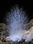
Autor: Andrea Massagli, Licencja: CC BY 4.0
The dimensions sometimes deceive. This is a little mildew (Gen. Mucor) found in a cave with some holes on the ceiling so the light can come inside. Colonies of this fungal genus grow fast, at rapid rate. They grow on organic matter in decomposition, on feces and other similar matter. It’s quite blue because I had a lamp whit a cold light, its real colour would be totally white with this singular quite transparent filaments. Mucor spores can be simple or branched and at the top have globular sporangia that are supported and elevated by a column-shaped columella. Only with my breath i could see the tiny filament move. (Cave “Buca delle Fate”, Asciano, Pisa, Italy)