Human brain right dissected lateral view description
Autor:
John A Beal, PhD
Dep't. of Cellular Biology & Anatomy, Louisiana State University Health Sciences Center Shreveport
Attribution:
Obraz jest oznaczony jako „Wymagane uznanie autorstwa” (attribution required), ale nie podano żadnych informacji o uznaniu autorstwa. Prawdopodobnie parametr atrybucji został pominięty podczas korzystania z szablonu MediaWiki dla licencji CC-BY. Autorzy mogą znaleźć tutaj przykład prawidłowego korzystania z szablonów.
Credit:
Krótki link:
źródło:
Wymiary:
653 x 413 Pixel (40769 Bytes)
Opis:
Human brain right dissected lateral view description.JPG
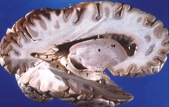
Lateral Portion of Frontal, Parietal, Occipital, and Superior Portion of Temporal Lobe Resected.
The anterior horn of the lateral ventricle is located in the frontal lobe. The body of the lateral ventricle continues posteriorly into the parietal lobe, the posterior horn into the occipital lobe, and the inferior horn down into the temporal lobe. Some structures produce elevations or bumps in the walls of the posterior and/or inferior horns of the lateral ventricles.
- Ventriculus lateralis, Cornu frontale
- Ventriculus lateralis, Pars centralis
- Calcar avis
- Ventriculus lateralis, Cornu occipitale
- Trigonum collaterale
- Eminentia collateralis
- Hippocampus
- Ventriculus lateralis, Cornu temporale
- Capsula interna
- Nucleus caudatus
Licencja:
Warunki licencji:
Creative Commons Attribution 2.5
Więcej informacji o licencji można znaleźć tutaj. Ostatnia aktualizacja: Fri, 16 Dec 2022 15:02:46 GMT