Myxomatous aortic valve
Autor:
Attribution:
Obraz jest oznaczony jako „Wymagane uznanie autorstwa” (attribution required), ale nie podano żadnych informacji o uznaniu autorstwa. Prawdopodobnie parametr atrybucji został pominięty podczas korzystania z szablonu MediaWiki dla licencji CC-BY. Autorzy mogą znaleźć tutaj przykład prawidłowego korzystania z szablonów.
Credit:
Praca własna
Krótki link:
źródło:
Wymiary:
2048 x 1536 Pixel (349803 Bytes)
Opis:
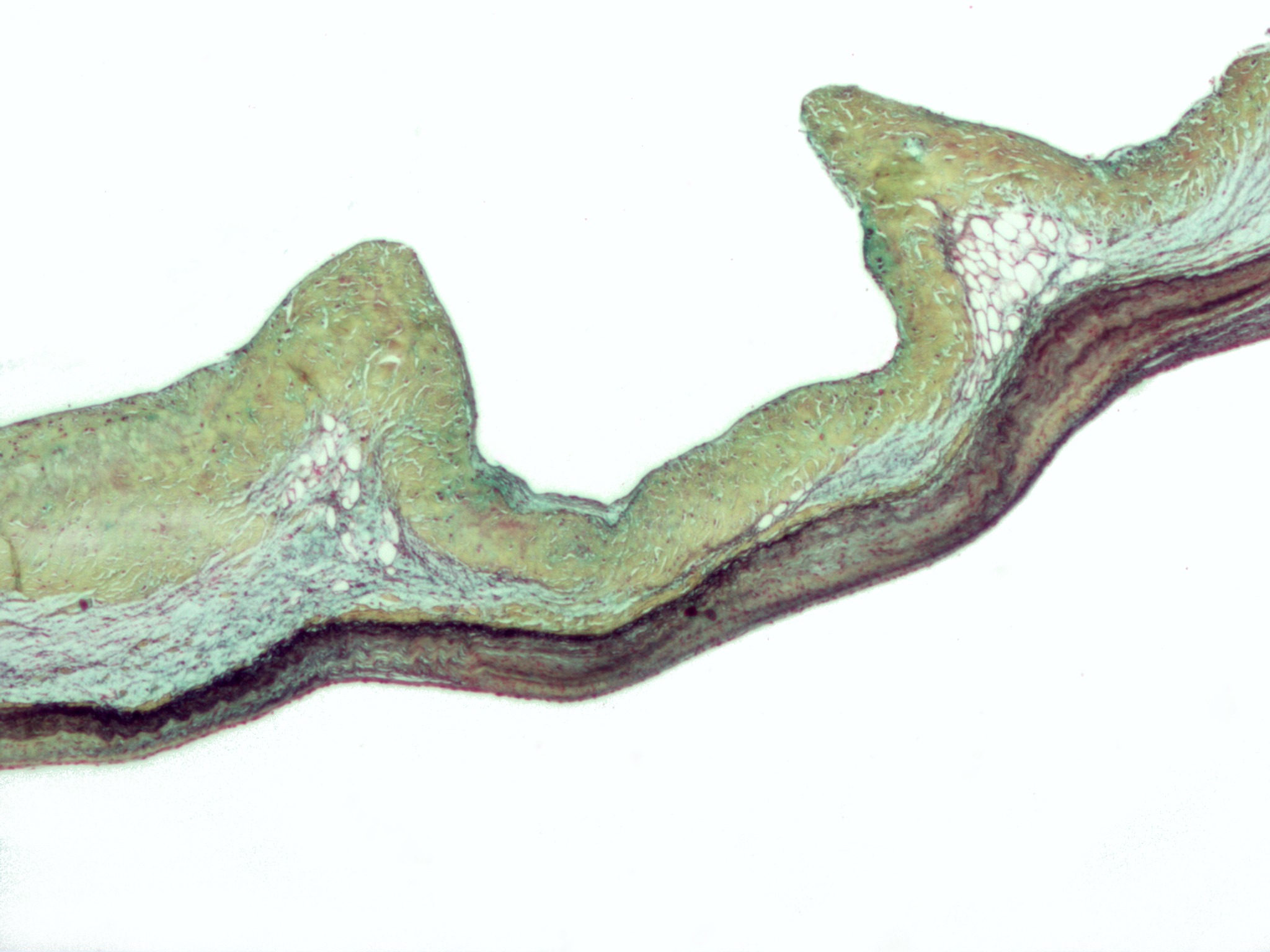
Micrograph of myxomatous degeneration of the aortic valve. Surgical specimen. Movat's stain (Black = nuclei, elastic fibres. Yellow = collagen, reticular fibers. Blue =
ground substance, mucin. Bright red = Fibrin. Red = muscle.)
In myxomatous degeneration, the ventricularis layer (composed primarily of elastic tissue) is thinned and the spongiosa layer (composed of loose connective tissue) is thickened.
On the image, the fibrosa layer (composed of collagen) is on the top, the thickened spongiosa layer below it and the ventricularis layer (made of elastic tissue) at the bottom.
The ventricularis layer, as the name may suggest, is closest to the (left) ventricle. The fibrosa layer is closest to the sinus of valsalva.
See also
- Marfan's syndrome - a condition, due to a defect in fibrillin (an essential component of elastic fibers), in which myxomatous degeneration is common.
Licencja:
Warunki licencji:
Creative Commons Attribution-Share Alike 3.0
Więcej informacji o licencji można znaleźć tutaj. Ostatnia aktualizacja: Thu, 24 Nov 2022 04:29:01 GMT