Thoracic anatomy
Autor:
- Patrick J. Lynch, medical illustrator. http://patricklynch.net
Yale University Center for Advanced Instructional Media - C. Carl Jaffe; MD; cardiologist
Attribution:
Obraz jest oznaczony jako „Wymagane uznanie autorstwa” (attribution required), ale nie podano żadnych informacji o uznaniu autorstwa. Prawdopodobnie parametr atrybucji został pominięty podczas korzystania z szablonu MediaWiki dla licencji CC-BY. Autorzy mogą znaleźć tutaj przykład prawidłowego korzystania z szablonów.
Credit:
Patrick J. Lynch, medical illustrator
Krótki link:
źródło:
Wymiary:
2304 x 1927 Pixel (1419935 Bytes)
Opis:
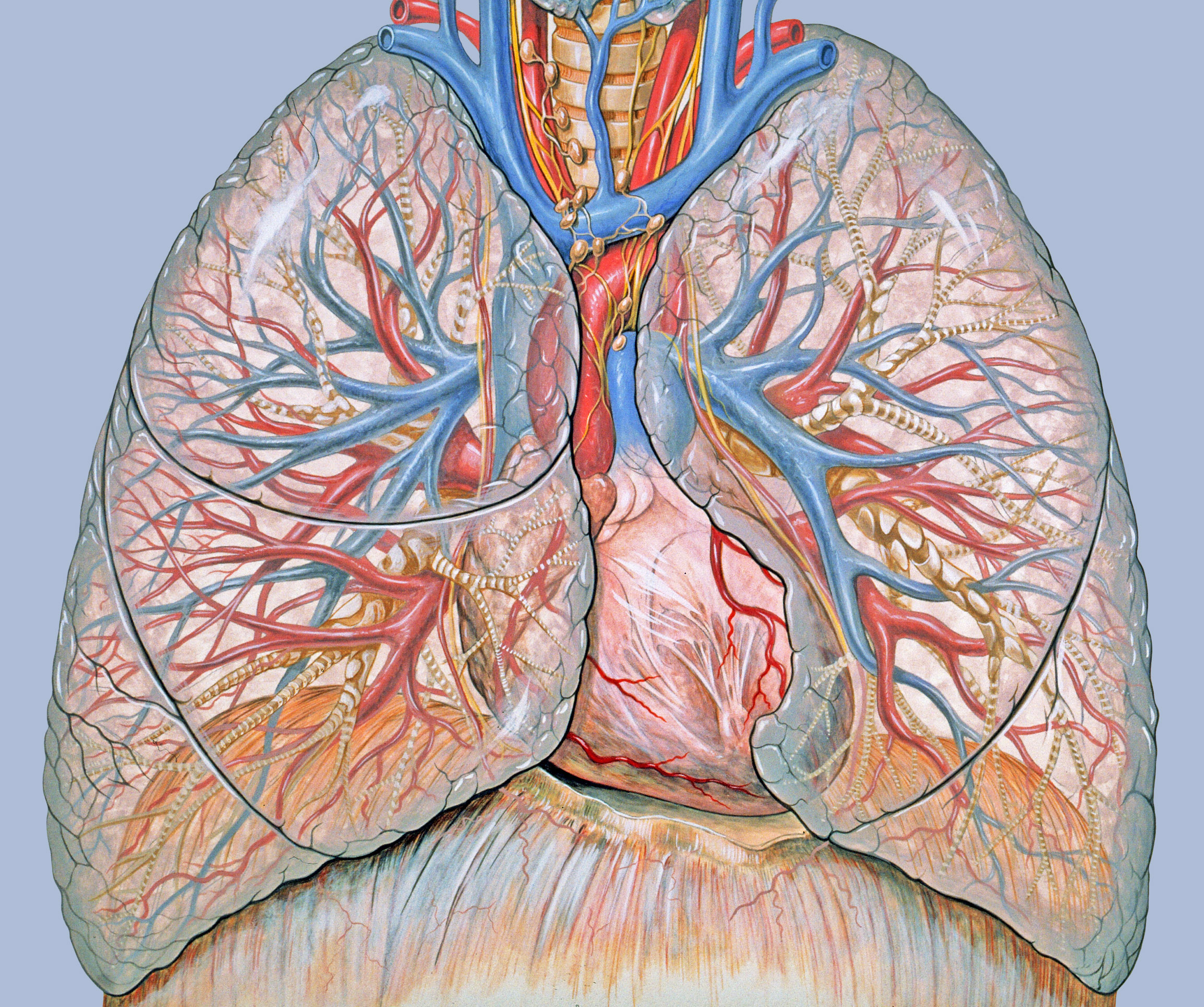
General thoracic anatomy, done specifically to support chest and heart imaging techniques. Generated for multimedia teaching projects by the Yale University School of Medicine, Center for Advanced Instructional Media, 1987-2000.
Errors
- Pulmonary arteries generally run alongside bronchi instead of being separate. (Pulmonary Artery Anatomy. University of Virginia (2013).)
- Each lobar vessel is in reality more clearly directed towards its corresponding lung lobe.
Keywords
chest, lungs, heart, trachea, anatomy, thoracic, thorax, pulmonary, anatomy, cardiac anatomy, diaphragm
Licencja:
Komentarz do licencji:
Creative Commons Attribution 2.5 License 2006
Warunki licencji:
Creative Commons Attribution 2.5
Więcej informacji o licencji można znaleźć tutaj. Ostatnia aktualizacja: Wed, 26 Oct 2022 19:24:03 GMT